Colour vision
Do you know if MS has affected you optic nerve? This is important to know if you want to assess your own EDSS.
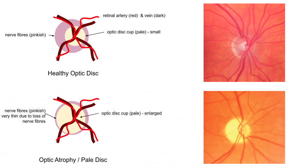
When I look in the eyes of my patients with MS using a device called an ophthalmoscope I am looking for the telltale signs of previous damage to the optic nerve. The sign we look for is optic disc pallor; the paleness of the so-called head of the optic nerve.
The optic disc is made up of nerve fibres from the retina, which then pass out of the back of the eye to form the optic nerve. If you have had optic neuritis in the past and have lost nerve fibres this can be detected with an ophthalmoscope, OCT (optical coherence tomography) or with retinal photography. Nerve fibre loss from optic neuritis makes the optic disc look pale (see figure below).
The optic disc receives its blood supply from small arteries from the back of the eye; the amount of blood is proportional to the number of nerve fibres in the optic disc. The lower the number of nerve fibres the fewer blood vessels there are the paler the disc looks. Please remember red blood cells are red and give a health optic disc a pinkish colour (see top images above).
Did you know that with a typical attack of optic neuritis you lose about 20% of the nerve fibres in the eye? If you lose so many nerve fibres why isn’t your vision so badly affected in that eye? That is simply because your visual system is able to compensate for the damage; it has spare capacity. Despite this most people with MS (pwMS) who have optic neuritis will know that although their visual acuity, or gross vision, may have recovered they have subtle deficits that we don’t routinely test for. For example, colour vision is often abnormal; colours appear washed out. Contrast sensitivity is also abnormal; you may have difficulty distinguishing between shades of grey. Your depth perception may also be affected; you need binocular (both eyes) vision for accurate depth perception. If you have poor depth perception you may see things in 3D when they should be in 2D and you may have difficulty judging distances. You may also find that you are hypersensitive to bright lights or lights with certain wavelengths; over the years I have noticed that a lot of pwMS become intolerant of fluorescent lights after an attack of optic neuritis.
The problem with remote consultations using video platforms, which I have used almost exclusively during the COVID-19, is that I can do this very important aspect of the neurological examination. Is it important? Yes, firstly it allows one to determine what neuronal systems have been affected by MS, which is required for diagnosis, i.e. dissemination of disease in space, and secondly it is important for assessing your EDSS or Expanded Disability Status Scale. The visual system is one of the functional systems in the EDSS and its involvement affects your EDSS.
When you use our web-EDSS online calculator it requires you to know if your neurological examination is normal or abnormal. Having optic disc pallor is not something you can get assessed remotely at present. This may change in the future; there are some very cool retinal cameras that clip onto your smartphone that may allow remote monitoring of retinal eye disease in the future.
However, you can get a reasonable idea if your optic nerve has been affected by MS by downloading and using one of the many colour vision applications on your smartphone; I recommend using ‘eye handbook’ as the basic version is free. Therefore, before completing your webEDSS I would check out your colour vision in both your eyes. This can be used as a proxy for optic nerve involvement. Please note if you have congenital colour blindness, which is more common in males, you can’t use this sign as a proxy for optic nerve involvement.
General Disclaimer: Please note that the opinions expressed here are those of Professor Giovannoni and do not necessarily reflect the positions of Barts and The London School of Medicine and Dentistry nor Barts Health NHS Trust. The advice is intended as general advice and should not be interpreted as being personal clinical advice. If you have problems please tell your own healthcare professional who will be able to help you.


My first symptom was ON and I definitely struggle with flashing or flickering lights. I don’t know whether it’s related but I find it difficult/impossible to appreciate the Op Art of Bridget Riley! My partner loves it and took me to the Tate Modern which turned out to be a nightmare. The patterns made me feel nauseous-not sure if this is an MS thing or not?
I've had five relapses, optic neuritis was the second. I have a pale optic disc as a result and definitely some sensitivity to fluorescent light ever since. Other bright lights and reflected light (eg on a window, mirror or screen) are hard to deal with too. My colour vision was fine even with the optic neuritis (as far as it could be tested - when it was bad I couldn't make out the largest letter on the eye chart so trying to pick the numbers in the colour vision test was impossible)