
Incidentalomas in Multiple Sclerosis
Every so often, a scan reveals something unrelated to your MS, such as a small cyst, a benign lump, a blood vessel variation, or a bit of inflammation in your sinuses.

What is an incidentaloma?
If you live with multiple sclerosis (MS), you will have many MRI scans over your lifetime; first to help make the diagnosis, and then at regular intervals to monitor your disease and check how well your treatment is working. MRI is a remarkably detailed way to look at the brain and spinal cord, and it is very good at detecting abnormalities.
Sometimes it picks up too much. Every so often, a scan reveals something unrelated to your MS, such as a small cyst, a benign lump, a blood vessel variation, or a bit of inflammation in your sinuses. Because these findings are discovered by chance rather than sought, doctors call them incidental findings, or, more informally, incidentalomas. The name simply means “a thing found incidentally”; it does not imply anything about its seriousness.
Incidentalomas are not unique to MS. They turn up whenever people have brain scans, whatever the reason. But people with MS are scanned far more often than the general population, which means they have more opportunities for one of these chance findings to appear.
How common are they?
More common than most people expect, but they are usually harmless.
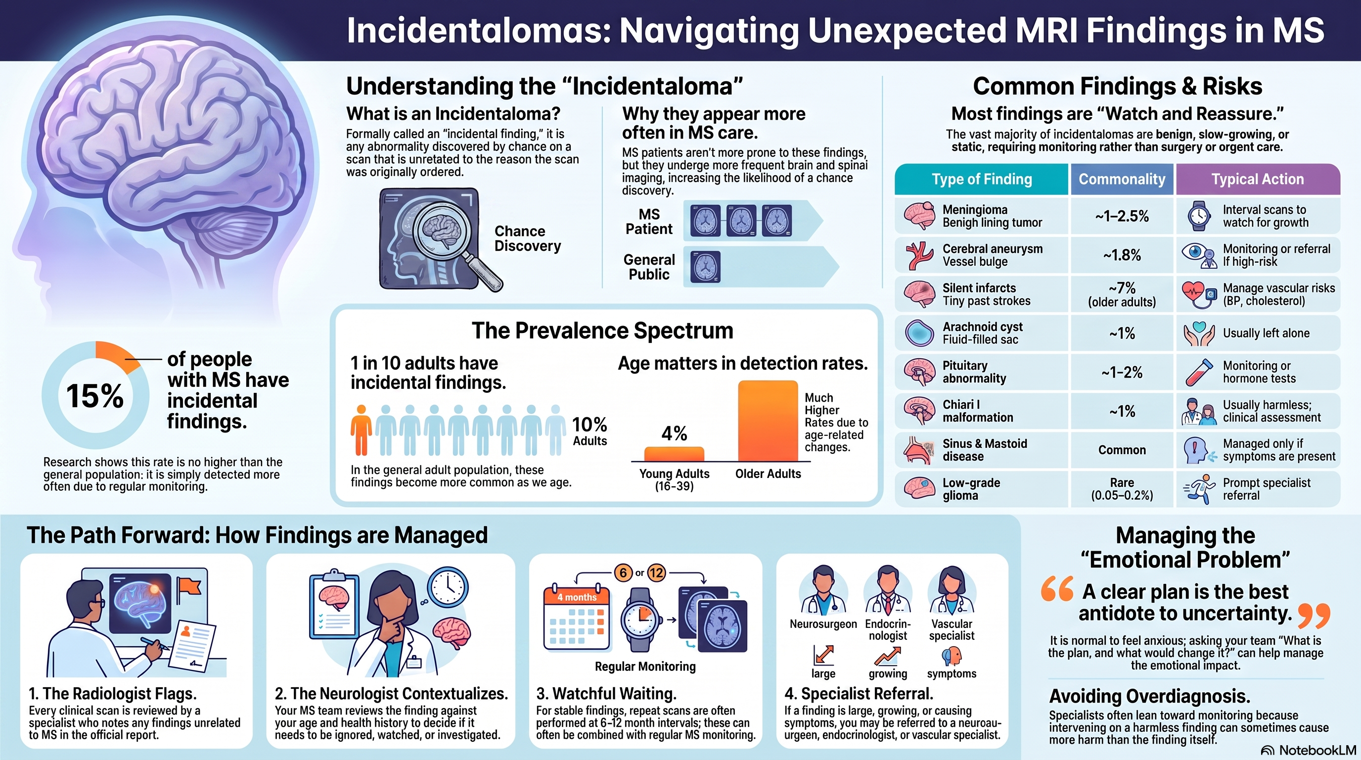
In the general adult population, large-scale studies provide a good baseline. According to research retrieved from PubMed and the wider literature, the population-based Rotterdam Scan Study found that roughly 1 in 10 adults has some incidental finding on a brain MRI, a figure that rises with age. The most frequent findings were silent (symptom-free) small strokes in about 7% of older adults, small brain aneurysms in about 1.8%, and benign tumours — mostly meningiomas — in about 1.6% (Please see paper 1 below).
In younger, healthy adults, the picture is reassuring. According to a large French student imaging study, only about 4% of people aged 18–35 had any incidental finding, and fewer than 2% had one that needed a referral to a specialist.
What about people who already have MS? A Canadian study that specifically looked at this question found incidental abnormalities in about 15% of people with MS, a rate no higher than in the matched comparison group without MS. The authors concluded that although MS itself does not create extra incidental findings, the sheer number of scans people with MS undergo means a sensible plan for handling these findings is essential (Paper 2 below).
The key message from these studies is that incidental findings are common, but serious ones are rare.
The spectrum of incidentalomas
Incidentalomas fall into a few broad groups. The table below summarises the most common types, roughly how often they occur, and what usually happens next. The percentages are drawn from general-population studies and will vary with age and scan quality.
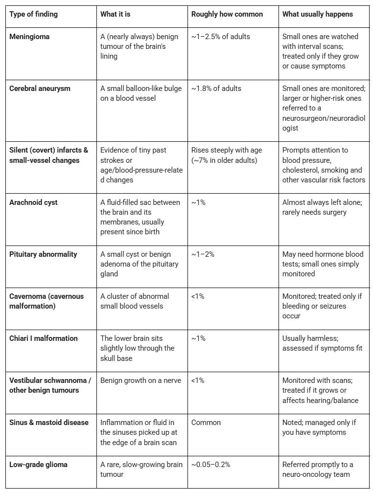
Most items on this list are benign, slow, or static. The purpose of listing them is not to alarm but to show that the vast majority sit in the “watch and reassure” category rather than the “urgent action” category.
A special case: findings that look like MS in people who don’t have symptoms
There is a mirror-image situation worth knowing about, especially for family members who may themselves have a scan one day. Occasionally, someone with no neurological symptoms has a brain MRI for an unrelated reason, for example, headaches or after a minor head injury, and the scan shows white matter spots that look exactly like MS. When there is no clinical history of MS, this is called radiologically isolated syndrome (RIS).
RIS is essentially “MS-looking findings found by accident.” Roughly 30–50% of people with RIS go on to develop MS within about five years, with the risk higher in younger people, men, and those with lesions in the spinal cord. For this reason, people with RIS are usually assessed by an MS specialist, may have additional spinal imaging and a lumbar puncture, and are followed closely. As you are now aware, many people with RIS can now be classified as having asymptomatic MS and in many situations be offered treatment. The treatment of RIS is not universal and often depends on local and national guidelines. On the NHS, for example, someone with RIS cannot be offered a DMT.
How incidentalomas are monitored and managed
When something unexpected appears on your scan, there is a well-trodden path for dealing with it, and it rarely involves anything dramatic:
1. The radiologist flags it. Every clinical scan is reviewed by a radiologist, who describes any finding unrelated to your MS in the report.
2. Your neurologist puts it in context. Your MS team looks at the finding alongside your age, symptoms, and general health, and decides whether it is a “leave alone,” “keep an eye on,” or “investigate” situation. A great many incidentalomas need nothing more than a note in your records.
3. “Watchful waiting” with interval scans. For findings such as small meningiomas, cavernomas, or aneurysms, the usual approach is a repeat scan at a set interval (often 6–12 months, then less frequently) to check whether anything has changed. Something that stays stable over time is very reassuring. Conveniently, people with MS are already having regular MRIs, so monitoring can often be folded into existing monitoring protocols.
4. Referral when warranted. If a finding is larger, growing, in a high-risk location, or causing symptoms, you will be referred to the relevant specialist, i.e., a neurosurgeon, a neuroradiologist for detailed vascular imaging, or an endocrinologist for a pituitary finding. Referral is a precaution, as the management of incidentalomas is usually handled by other subspecialists.
5. Attention to modifiable risk. Some findings, especially silent small strokes and small-vessel changes, are less about a single lesion and more a nudge to look after your blood pressure, cholesterol, blood sugar, weight, and to stop smoking. These same steps are good for your brain health with MS in any case.
The part nobody warns you about: the worry
An incidentaloma is often more of an emotional problem than a medical one. Being told there is “something else” on your brain scan is unsettling, particularly when you are already coping with MS. It is completely normal to feel anxious while you wait for an explanation or a follow-up scan.
Two things help. First, remember the numbers: most incidental findings are benign and never cause trouble. Second, ask questions. It is entirely reasonable to ask your MS team, “What exactly was found? How serious is it? What is the plan, and what would change it?” A clear plan is the best antidote to uncertainty. Some findings carry a small risk of “overdiagnosis” and “overmanagement”, i.e., being investigated or treated for something that would never have harmed you, which is exactly why specialists lean towards careful monitoring rather than intervening with most incidentalomas.
The bottom line
Incidental findings on MRI are a by-product of good MS care. Because you are scanned regularly, the odds are reasonable that at some point a scan will show something unrelated to your MS. In the overwhelming majority of cases, that “something” is harmless, slow-moving, or easily monitored, and it changes nothing about your MS treatment. A small minority need specialist input, and a well-organised MS service is set up to sort one from the other.
If you receive a report mentioning an incidental finding, try not to jump to conclusions. Bring it to your MS team, ask for the plan in plain language, and let the monitoring and specialists do their job. More often than not, the story ends with a stable follow-up scan and a note in your file.
I would be interested to know if any of you have had incidentalomas found on your MRI. What was the diagnosis, and how has it been managed?
Paper 1
Background: Magnetic resonance imaging (MRI) of the brain is increasingly used both in research and in clinical medicine, and scanner hardware and MRI sequences are continually being improved. These advances are likely to result in the detection of unexpected, asymptomatic brain abnormalities, such as brain tumors, aneurysms, and subclinical vascular pathologic changes. We conducted a study to determine the prevalence of such incidental brain findings in the general population.
Methods: The subjects were 2000 persons (mean age, 63.3 years; range, 45.7 to 96.7) from the population-based Rotterdam Study in whom high-resolution, structural brain MRI (1.5 T) was performed according to a standardized protocol. Two trained reviewers recorded all brain abnormalities, including asymptomatic brain infarcts. The volume of white-matter lesions was quantified in milliliters with the use of automated postprocessing techniques. Two experienced neuroradiologists reviewed all incidental findings. All diagnoses were based on MRI findings, and additional histologic confirmation was not obtained.
Results: Asymptomatic brain infarcts were present in 145 persons (7.2%). Among findings other than infarcts, cerebral aneurysms (1.8%) and benign primary tumors (1.6%), mainly meningiomas, were the most frequent. The prevalence of asymptomatic brain infarcts and meningiomas increased with age, as did the volume of white-matter lesions, whereas aneurysms showed no age-related increase in prevalence.
Conclusions: Incidental brain findings on MRI, including subclinical vascular pathologic changes, are common in the general population. The most frequent are brain infarcts, followed by cerebral aneurysms and benign primary tumors. Information on the natural course of these lesions is needed to inform clinical management.
Paper 2
Background: Incidental findings arising from imaging research have important implications for patient safety. Magnetic resonance imaging is widespread in multiple sclerosis (MS) studies and care, yet the prevalence rate of incidental findings in MS is poorly defined. The absence of such reports in the MS literature suggests that such findings may be deemed inappropriate for documentation in research publications, or possibly, not fully reported at all.
Objective: We sought to document incidental findings from a study designed to detect features of chronic cerebrospinal venous insufficiency (CCSVI) in MS patients and control subjects.
Methods: Magnetic resonance images were obtained as part of a prospective study conducted between October 2010 and September 2012. Patients with MS (relapsing-remitting, primary progressive, secondary progressive), clinically isolated syndromes, and neuromyelitis optica and age/sex-matched healthy controls were included. All images were reviewed by neuro-radiologists for quality-control purposes.
Results: Magnetic resonance imaging was successfully obtained in 166 participants (110 patients, 56 controls). Incidental abnormalities (n = 33) were detected in 15% of patients (n = 17) and 27% of controls (n = 15), comprising 19% overall (n = 32).
Conclusions: The prevalence of incidental findings from the MS population was not significantly different from the control population. However, the overall prevalence was high and warrants a careful management strategy for future imaging studies.
Accidental readers
If you have been forwarded this email and are not an MS-Selfie subscriber, please consider subscribing and helping MS-Selfie expand its resources for the broader MS community. MS-Selfie relies on subscriptions to fund its curated MS-Selfie microsite, MS-Selfie books, MS-Selfie Infocards, and other activities that extend beyond the MS-Selfie Substack newsletters.
Subscriptions and donations
MS-Selfie newsletters and access to the MS-Selfie microsite are free. In comparison, off-topic Q&A sessions are restricted to paying subscribers. Subscriptions are being used to run and maintain the MS Selfie microsite and other related activities, as I don’t have time to do this myself. You must be a paying subscriber to ask questions unrelated to the newsletters or podcasts. If you can’t afford to become a paying subscriber, please email a request for a complimentary subscription (ms-selfie@giovannoni.net).
Questions
If you have questions unrelated to the newsletters or podcasts, please email them to ms-selfie@giovannoni.net. Prof. G will try to answer them as quickly as possible.
Important Links
🖋 Medium
General Disclaimer
Please note that the opinions expressed here are those of Professor Giovannoni and do not necessarily reflect the positions of Queen Mary University of London or Barts Health NHS Trust. The advice is intended as general and should not be interpreted as personal clinical advice. If you have any problems, please tell your healthcare professional, who can help you.
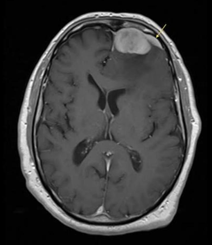
Yep, this is me. Incidental small meningioma picked up on MRI in 2023 (possibly there since 2018). Watch & Wait for a year and then a year of it growing on two consecutive six month scans so surgery was recommended whilst it was still relatively small and uncomplicated. Had the surgery a couple of months ago. I was referred to a neurosurgeon as soon as the meningioma was confirmed and he took over care and action for it, whilst my neurologist continued to manage my MS care but also offered advice too. Fantastic neurosurgical team who were always cognizant of my MS throughout. I completely agree with the emotional impact. One of my biggest worries was how surgery to the brain might affect my MS. Also my neurosurgeon said that radiation wouldn't be recommended for me because of my MS. Conversely, am thankful for my MS because without it I probably wouldn't have known about it until the meningioma had started to cause symptoms and been much bigger. Also my neurosurgeon isn't worried about ongoing monitoring post-surgery because we're already doing MRIs because of my MS. I've also come across a few others like me who have MS and who have been diagnosed with meningiomas.
You remind me of the century of MS autopsies carried out on MS cases, throwing away meningeal tissues (both leptomeningeal and dural) with 'inflammation, as 'incidental', 'accidental' or irrelevant. Stephen Hauser makes the point in his lovely book ('The face laughs while the Brain Cries'). These, his mentors assured him to be 'postmortem artefacts' 'This inflammation has nothing to do with MS. It is seen in every brain regardless of the cause of death' The wisdom of experts. Despite the most detailed observations of James Dawson,(1916!!) 'these meningeal changes are 'of the greatest importance in relation to the pathogenesis of disseminated sclerosis'. Meningeal inflammations and their topography have suddenly become the 'flavour of the moment' and the clever explanation of 'progression', the Real MS'
You can see therefore my irritation when 40 years worth of publications to try to stimulate action on the remarkable associations and significant similarities between paranasal sinus mucosal inflammations and MS, virtually ignored. Not least irritated when you classify the commonest 'incidental' finding in MS cases as "Inflammation or fluid of the sinuses picked up at the edge of a brain scan'' I wish I had stuck to my guns' writes Stephen Hauser.....but I didn't due to my respect for the wisdom of experts' I think I will stick to my guns. If at all interested Mr Google will provide the published data.