ACTRIMS 2025: How old is your brain?
As brain age can be relatively easily measured using MRI and AI, would you want to know the age of your brain relative to your chronological age?
One of the main themes covered at ACTRIMS this year was MS and ageing.
We have known for some time that brain aging is accelerated in MS and that it is associated with worse physical and cognitive disability and more significant change in disability over time. I have always included brain ageing as one of the drivers of smouldering MS. I suspect brain age is an integrator at the level of the end-organ of all the damage that accumulates over time.
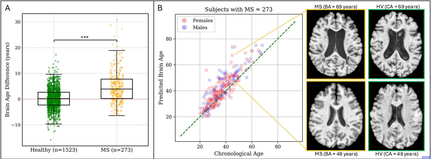
Erin Beck from Mount Sinai (New York) gave an excellent presentation on using MRI to determine brain age. She showed that accelerated brain ageing is more pronounced in early MS. She reviewed the evidence that accelerated brain aging is associated with physical and cognitive disability and a more significant change in disability over time. Of interest is that she uses an AI method to analyse the MRI of the brain that generates a single metric to represent brain age. On average, people with MS have a brain age that is approximately 10 years older than their chronological age.
She presented evidence that focal (lesions) and diffuse (smouldering) brain damage contribute to brain ageing. She implied that brain age could be used prognostically and potentially as a biomarker of a treatment response in MS. If a particular MS therapy works, it should slow down the premature accelerated brain ageing that is seen in MS. Or even better, can we reverse the brain age signature with neurorestorative therapies?

In parallel, a fascinating paper (see paper below) has just been published, showing an increase in somatic single-nucleotide variants (sSNVs) in neurons from MS lesions compared to normal-appearing MS and control tissues. These sSNVs are somatic mutations in the DNA of individual neurons that are not present in the so-called inherited germline DNA. The investigators found an increase of ~44 mutations per year in neurons from chronic MS lesions, which is 2.5 times faster than in neurons from normal-appearing MS and control tissues. A signature analysis of the mutations suggested that the mutations were age-associated and probably due to neuroinflammation.
These two pieces of evidence support other findings that inflammation harms you and causes premature ageing, which targets the brain in the case of MS. We need proof that treating MS early and effectively stops or prevents premature ageing. I suspect the data using MRI brain age is already out there. We only need several pharmaceutical companies to analyse the trajectory of MRI brain ageing in the subjects in their clinical trials. I would predict that subjects randomised to highly effective DMTs would do better than subjects randomised to placebo or lower efficacy DMTs.
As brain age can be relatively easily measured using MRI and AI, would you want to know the age of your brain relative to your chronological age? If you are 40 years old and your brain age comes back as 52, i.e. 12 years older than your chronological age, how would you use this knowledge?
Abstract 1
E. Beck. MRI Assessment of Biological Age in MS. S2.1. ACTRIMS 2025
There is a complex interplay between aging and most aspects of MS pathology that are visualized on MRI, including focal lesion formation, chronic inflammation, and repair, as well as more diffuse structural changes. MS may potentiate age-related changes in the immune and central nervous systems, leading to accelerated aging and increased chronic inflammation, decreased repair, and increased diffuse myelin and neuro-axonal damage with age, ultimately resulting in more rapidly progressive disability. In young people, new focal inflammatory demyelination in both the white and gray matter is more common, but new lesions are more likely to repair and less likely to demonstrate chronic inflammation than in older people. To better understand how aging and MS interact to yield more diffuse structural changes in the brain, we use large MRI datasets of healthy individuals and both statistical and deep learning-based techniques to model changes in individual structural measures, including brain volume and cortical curvedness, as well as brain age, a more global measure derived directly from T1-weighted MRI, over the human lifespan. These models allow us to begin to untangle MS vs age-related changes in the brain and determine how brain aging influences the disease course. We find that brain aging is accelerated in MS, with more pronounced acceleration in early disease. Accelerated brain aging is in turn associated with worse physical and cognitive disability and greater change in disability over time. A better understanding of how both focal and diffuse central nervous system damage is both caused by and may contribute to aging is important for predicting outcomes in MS and targeting treatments toward aging-related processes, which may be important drivers of disability progression.
Article
Neuroinflammation underpins neurodegeneration and clinical progression in multiple sclerosis (MS), but knowledge of processes linking these disease mechanisms remains incomplete. Here we investigated somatic single-nucleotide variants (sSNVs) in the genomes of 106 single neurons from post-mortem brain tissue of ten MS cases and 16 controls to determine whether somatic mutagenesis is involved. We observed an increase of 43.9 sSNVs per year in neurons from chronic MS lesions, a 2.5 times faster rate than in neurons from normal-appearing MS and control tissues. This difference was equivalent to 1,291 excess sSNVs in lesion neurons at 70 years of age compared to controls. We performed mutational signature analysis to investigate mechanisms underlying neuronal sSNVs and identified a signature characteristic of lesions with a strong, age-associated contribution to sSNV counts. This research suggests that neuroinflammation is mutagenic in the MS brain, potentially contributing to disease progression.
Subscriptions and donations
MS-Selfie newsletters and access to the MS-Selfie microsite are free. In comparison, weekly off-topic Q&A sessions are restricted to paying subscribers. Subscriptions are being used to run and maintain the MS Selfie microsite, as I don’t have time to do it myself. You must be a paying subscriber if people want to ask questions unrelated to the Newsletters or Podcasts. If you can’t afford to become a paying subscriber, please email a request for a complimentary subscription (ms-selfie@giovannoni.net).
Important Links
🖋 Medium
General Disclaimer
Please note that the opinions expressed here are those of Professor Giovannoni and do not necessarily reflect the positions of Queen Mary University of London or Barts Health NHS Trust. The advice is intended as general and should not be interpreted as personal clinical advice. If you have problems, please tell your healthcare professional, who will be able to help you.


It would help determine whether the crushing fatigue, inability to focus, and significantly reduced resilience stems from or is more likely related to MS pathology -- as opposed to depression, stress, or laziness!
I've tried all available stimulants, many anti-depressants, ALCAR, B12, Nicotine, exercise, and meditation -- often in concert -- with little to no improvement.
I enrolled in law school 8 years after diagnosis and graduated near the top of my class. 6-7 years later, I am failing at a law adjacent job I used to crush with minimal effort. The resulting financial stress, compounded with a recent divorce leave me oscillating between anger, grief, and disappointment!
Untangling MS pathology from moral failings and/or other issues would help me unclench!
Very interesting article Gavin. I have some recent experience of a similar MRI/AI assessment called NeuroQuant. It provides the volume of specific brain structures affected by neurodegenerative diseases and compares these measurements to healthy brains of similar age, sex, and skull size. My brain is on the 1st percentile for size and the 98th for white matter hypointensities, 99th percentile for ventricle volume. 1st percentile for cerebral white matter and ventral diencephalon and 1st percentile for frontal cortex volume. I can see by looking at my MRI that my brain age is well above my chronological age. I had always pointed out the atrophy to my neurologists but they didn’t seem to have any helpful comments. In fact one neurologist told me “by the time he’d be worried about atrophy, I wouldn’t be.” I was expecting the results to be challenging which they were but also they were validating. As to what to do with the results… my GP has told me not to focus on it, however, it is worrying as I’m 48 and my brain looks more like 68. It has made me reevaluate what I am doing and made me look further into brain health. I’m on Natalizumab 6 weekly and my MRI is stable. I am trying to increase my activity levels and loose weight. I have always loved learning so I am considering doing some post-graduate courses in the hope that more brain connections equals a healthier brain. I’d still like some more direction on what other things I should be doing. I want to preserve what I’ve still got! Thank you for raising this issue. Jane